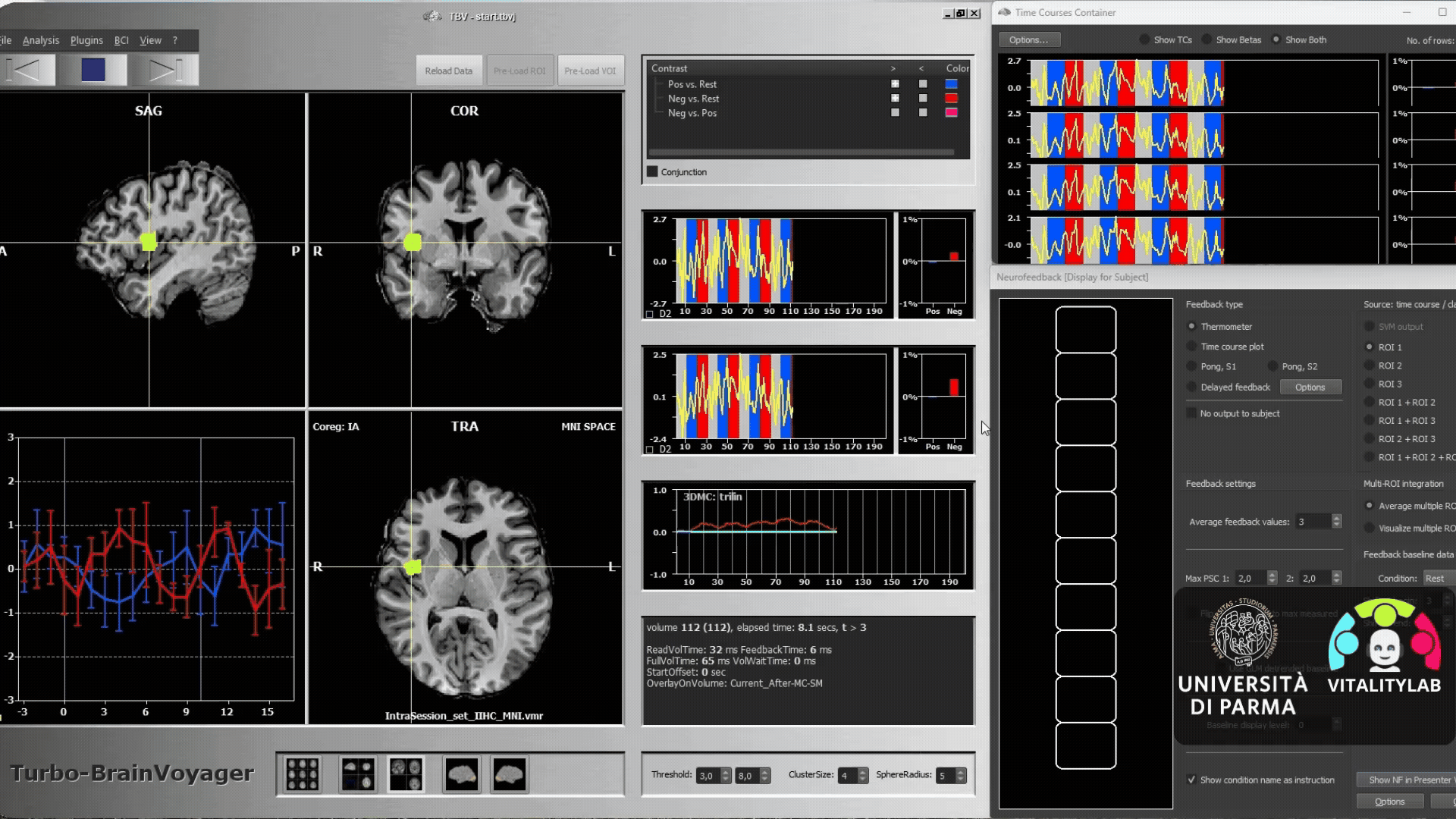
Functional magnetic resonance imaging (fMRI) is a non-invasive neuroimaging technique that indirectly measures brain activity. The subject is placed inside the resonance scanner where he or she must observe stimuli or perform tasks. The machine detects changes in blood oxygenation levels (BOLD signal) associated with neural activity. When brain regions are activated, they require increased oxygenated blood flow, which fMRI can measure. By identifying these regions, researchers can infer the neural correlates of various cognitive functions.
Let's take an example, in 2015 the group of Di Cesare and colleagues hypothesized that the dorso-central insula is involved in the encoding of vitality forms.
If you want to read more about this topic, click here Article PDF
Di Cesare, G., Di Dio, C., Marchi, M., & Rizzolatti, G. (2015). Expressing our internal states and understanding those of others. Proceedings of the National Academy of Sciences, 112(33), 10331-10335.
Through kinematics, we can calculate numerous parameters of movement, such as maximum acceleration, speed, and finger aperture, using markers placed on the right hand and infrared cameras. This technique has been employed in many experimental paradigms to assess the modulation of Vitality Forms in ethological contexts, measuring the position of the hand in X/Y/Z coordinates at each moment in time.
Here is an example of an experimental kinematics paradigm, in which a video clip is shown where an actor passes a bottle to the participant, or asks the participant to pass it to him. The kinematic and sound waves, relating to the actor's requests, are highlighted in red and blue. (Di Cesare et al., 2017)
If you want to read more about this topic, click here Article PDF
Di Cesare G., De Stefani E., Gentilucci, and De Marco D. (2017). Vitality Forms Expressed by Others Modulate Our Own Motor Response: A Kinematic Study. Front. Hum. Neurosci. 11:565.
Psychophysics is the scientific study of the relationship between physical stimuli from the external world and the subjective experiences these stimuli provoke in us. Some of these parameters are the estimation times, absolute threshold, the differential threshold, the sensory acuity, the reaction time, etc. In particular, in the field of neuroscience, it allows us to understand the mechanisms underlying perception. For example, Di Cesare et al. (2021) demonstrated that vocal requests conveying different vitality forms can influence the perception of goal-directed actions, affecting the estimation of action duration. Participants first heard a vocal request (‘give me’) in a rude or gentle tone, then had to observe the initial part of a rude or gentle passing action, continue it mentally and estimate the time of its completion. The results showed that listening to a gentle vocal request increased the estimated duration of an action, while listening to a rude vocal request decreased it. This contagion effect lasts 800 ms and then starts to decay.
Graphs representing the estimated timing of gentle (A) and rude (B) actions. Green bars refer to incongruent conditions: rude vocal request and gentle action (RDV_GTA) in panel (A), gentle vocal request and rude action (GTV_RDA) in panel (B). Blue bars in panel (A) refer to gentle congruent condition: gentle vocal request and gentle action (GTV_GTA). Red bars in panel (B) refer to rude congruent condition: rude vocal request and rude action (RDV_RDA). The graphs below show the effect of rude vocal requests on gentle action estimation (C) and the effect of gentle vocal requests on rude action estimation (D) for five different time delays (0, 400, 800, 1,200, and 1,600 ms). **p < 0.01, and ***p < 0.001. (Di Cesare et al. 2021)
If you want to read more about this topic, click here Article PDF
Di Cesare, G., Pelosi, A., Aresta, S. M., Lombardi, G., & Sciutti, A. (2021). Affective contagion: How attitudes expressed by others influence our perception of actions. Frontiers in Human Neuroscience, 15, 712550.
Physiological techniques utilize a range of methodologies and instrumentation to measure and analyze physiological variables, providing quantitative data on functions . Some of the techniques we are currently using are the electrocardiogram (ECG), to record the electrical activity of the heart, and electromyography (EMG), to record the electrical activity of muscles. The use of ECG in the field of vitality forms is currently under development while the application of the EMG technique can be seen in the study conducted by Rizzolatti et al. (2021). This work has shown that a handshake made in a rude or gentle manner induces increased activation of the dorso-central insula. Electromyographic recording was used to profile muscle responses.
The first graph (A1) shows the mean response profiles of the handshake in the aggressive, gentle or neutral condition, while the second graph (A2) shows the mean intensity, both obtained by EMG. The other two graphs show the mean response profile (B1) and mean response intensity (B2) during a neutral handshake and the observation of angry, happy and neutral facial expressions (Rizzolatti et al. 2021)
If you want to read more about this topic, click here Article PDF
Rizzolatti, G., D’Alessio, A., Marchi, M., & Di Cesare, G. (2021). The neural bases of tactile vitality forms and their modulation by social context. Scientific Reports, 11(1), 9095.